baby ultrasound pictures 4 weeks
Twin development at week 20. Folic acid greatly reduces your babys risk of developing neural tube birth defects such as spina bifida.
Ultrasound Scan Of A Child 20 Weeks 5 Month Pregnant Concept Stock Photo Alamy
By next month your babys lungs will be fully formed.
. Our content is doctor approved and evidence based and our community is moderated lively and welcoming. At 4 weeks pregnant the tiny life inside you which is technically an embryo is implanting in your uterus where it will grow and develop over the next 36 weeks. If youre interested in 3D ultrasound pictures try asking at your providers office.
Along with implantation in the uterine lining comes a rise in the pregnancy hormone hCG. In the next few weeks your baby will double his weight and add inches to his length. Her baby was on a ventilator for 48 hours and a feeding tube for six days.
Video technology allows Childrens Minnesota specialists to help save Wisconsin baby For most of us technology provides convenience. Babys Development at 24 Weeks. With your baby now weighing a little over 4 pounds you might be waddling and having trouble.
200 baby names that start with C Popular choices for baby names that start with C include classics like Christopher and Claire as well as more modern names like Cam and Casey. Baby development at 34 weeks Growing nails. For optimal facial 3D scan images we recommend that this is carried out between 26 to 32 weeks.
Shown here is a 2D ultrasound inset contrasted with a 4D ultrasound both at 20 weeks. School of Nursing Human Health Building Room 3027 433 Meadow Brook Road Rochester MI 48309-4452. Your baby is about the size of a sesame seed.
The iron stores your baby builds now last for the first 6 months of life until your little one starts eating solid foods. In some cases you may need an ultrasound at 34 weeks to check on your babys health or progress. Obstetrics Gynecology 646.
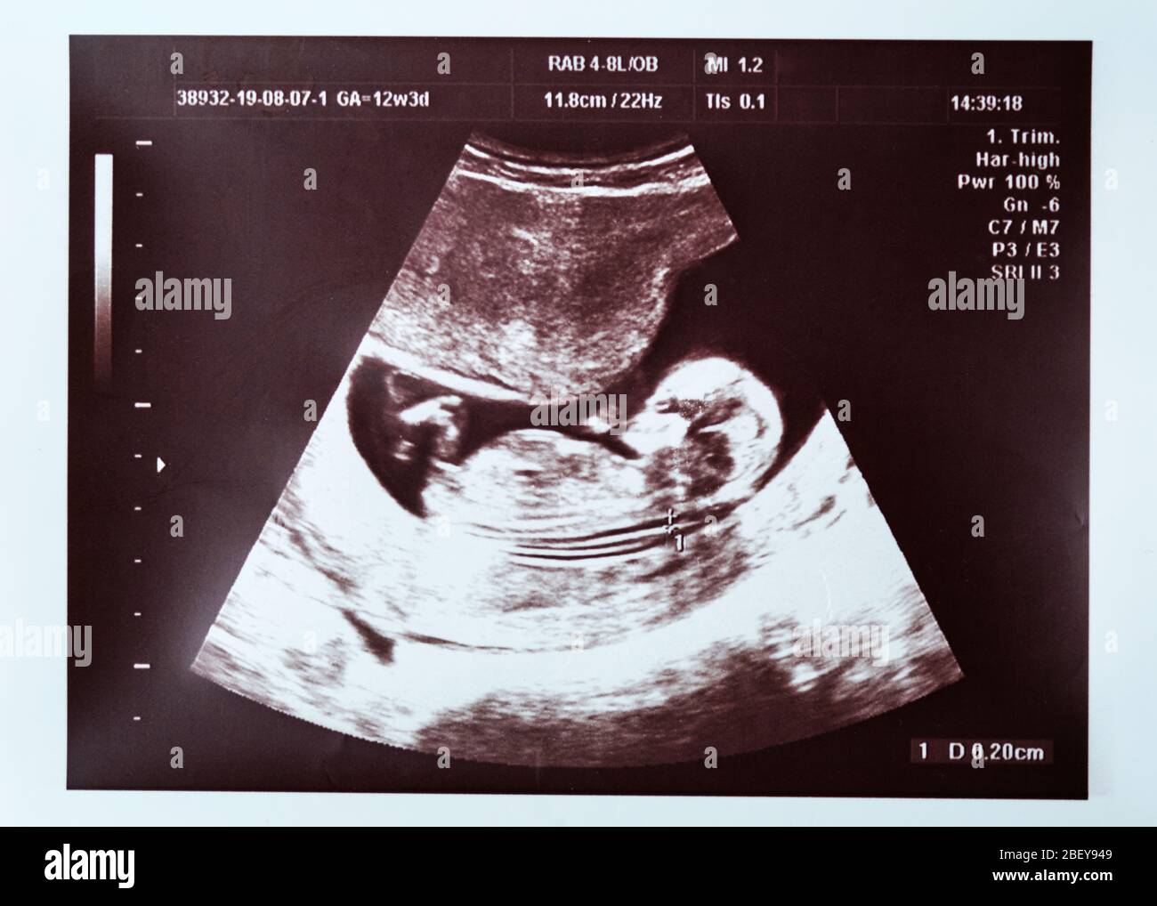
For example if you are having an ultrasound of your uterus then what you see at the top of the screen or printed ultrasound. If you havent started taking a prenatal vitamin yet nows the time to start. You need about 450 extra calories per day to support your pregnancy here are some healthy.
You likely had an ultrasound in your first trimester and wont have one again until the anatomy scan aka the mid-pregnancy ultrasound which usually happens between weeks 18 and 22The doctor will also use an ultrasound if you plan to have an amniocentesis between weeks 15 and 20At an. BabyCenter is committed to providing the most helpful and trustworthy pregnancy and parenting information in the world. Most practitioners wait until at least 6 weeks to perform the first pregnancy ultrasound.
Doppler studies show the sound response by measuring movement and heartbeat rates. If they have the equipment they may be able to provide you with a few pictures during a regular scan or schedule an additional one at an out-of-pocket cost. In the third trimester aim for a steady weight gain of about a pound each week.
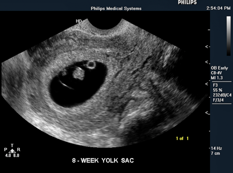
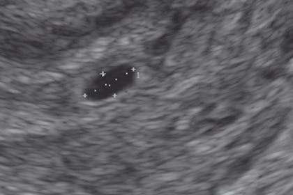
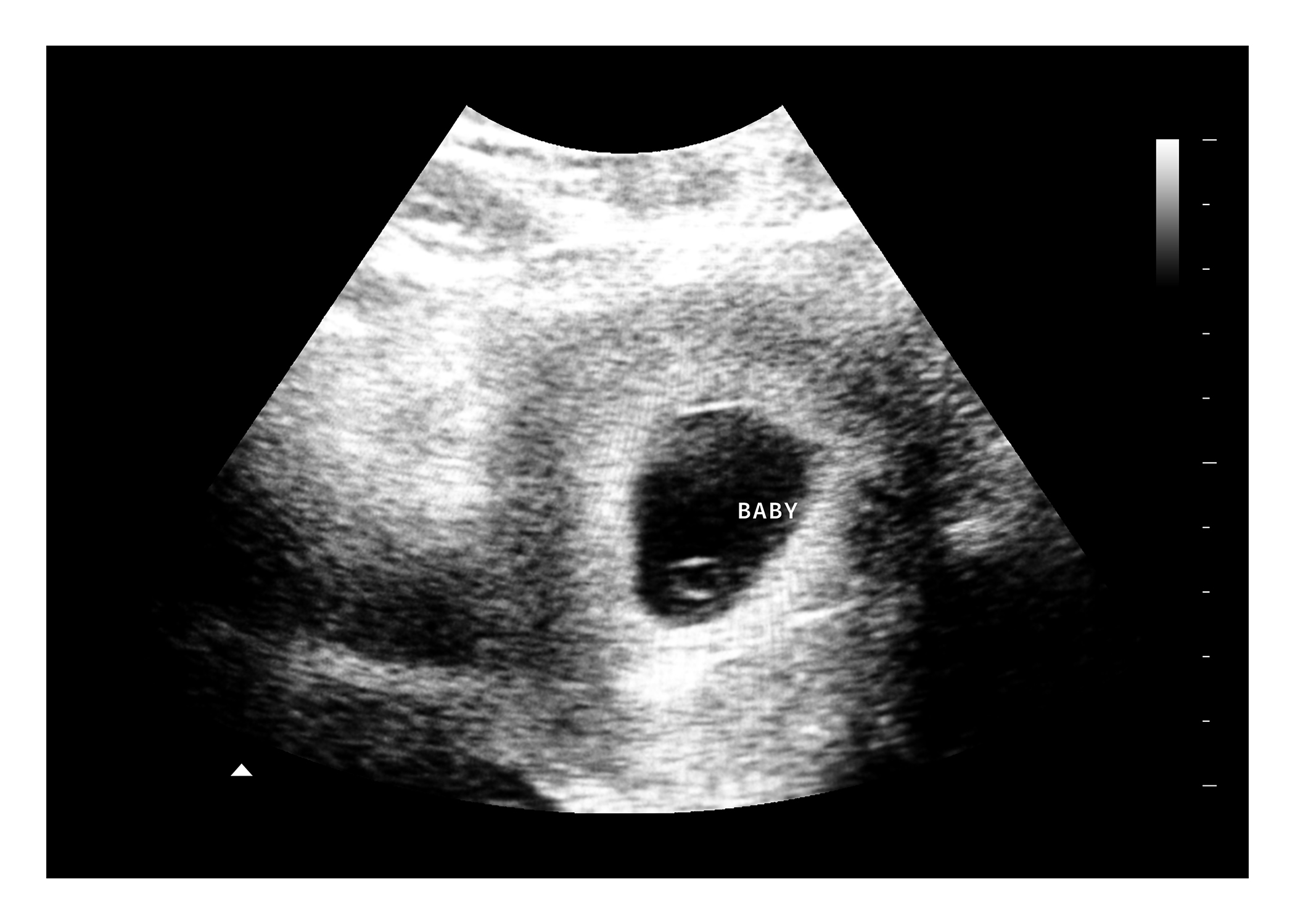
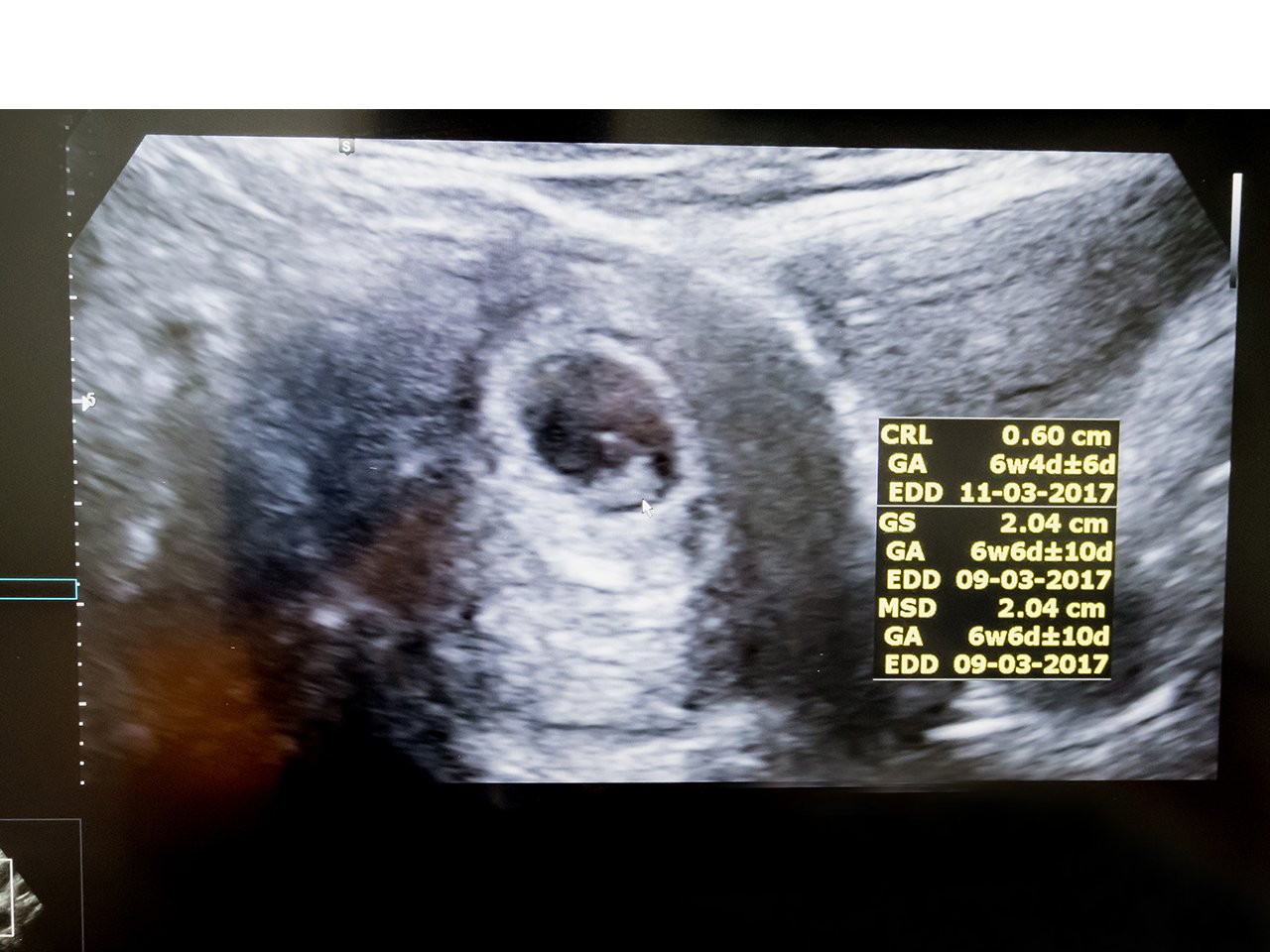
At 15 weeks pregnant your baby is nearly 4 12 inches CRL nearly 6 12 inches in height and weighs about 4 ounces. With thousands of award-winning articles and community groups you can track your pregnancy and babys growth get answers to your. However a gestational sac can be seen as early as 4 12 weeks after your last period and a fetal heartbeat can be detected at 5 to 6 weeks though that isnt always the case.
Shown here is a 2D ultrasound inset contrasted with a 4D ultrasound both at 20 weeks. Its particularly critical to get enough folic acid while trying to conceive and during your first trimester. Development at 24 Weeks.
According to a 2012 study published in the journal Obstetrics and Gynecology no less than 69 of parents wanted to know. Read more about whats happening at 4 weeks pregnant. The top of the screen or printed image is where the ultrasound probe was placed.
In 1995 a pregnant African-American doctoral student had a preterm birth after her water broke unexpectedly at 34 weeks. For this test the transducer is. Take a prenatal vitamin.
Your babys body is stashing away important minerals such as iron calcium and phosphorus. In the first trimester up to eight and rarely nine weeks the baby may not be seen by an ultrasoundThe abdominal scan may miss it a. 19 weeks pregnant.
The accuracy can vary from 703 at 11 weeks to 987 at 12 weeks and 100 at 13 weeksEleven weeks is the earliest that sex determination canNo they cant. Sometimes the baby will develop hiccups that the mother can feel. The baby weighs almost 4 pounds and is moving around often.
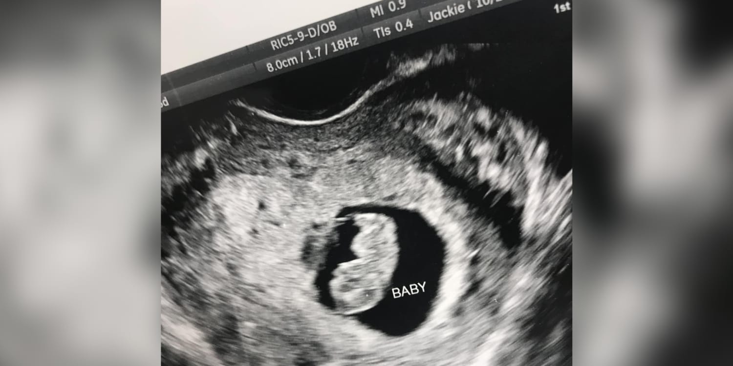
During the scan youll be able to hear your babys heartbeat during the ultrasound and youll go home with a few pictures of your baby. At 24 weeks the baby may weigh 14 pounds and can respond to sounds. Hello third trimester.
The ultrasound technician will put gel on your neck where your carotid arteries are located. You may have your first ultrasound early in pregnancy a first-trimester ultrasound or you may have a standard ultrasound at 18 to 22 weeks. Baby development at 32 weeks Storing minerals.
Your pregnancy may be detectable in a week or so by a home pregnancy test. The ultrasound femur length as a predictor of fetal length. Pregnancy ultrasound allows your provider to check on your babys health and development monitor your pregnancy and look for any physical abnormalities.
Hang pictures or do anything more than necessary to get through the day. They are the same type of sound waves that doctors use to create and record pictures of a baby inside a pregnant woman. Start from the top of the image.
Your babys genitals are developed enough to see on an ultrasound. In other words the image you see shows what the organ or tissues look like from the side rather than from the top. Typically there isnt a 14-week ultrasound.
One of the most common ways to do this is with an ultrasound most frequently performed at between 18 and 20 weeks of gestation. Learn more about your symptoms and how your baby is growing this week. At 34 weeks.
The uterus is not very large and the baby takes up basically all of it. Optional Add one of our heartbeat bears to this package for 20. Youre in the home stretch.
An increase in appetite is normal now. But for a western Wisconsin family technology was a true. Baby Scanning pregnancy ultrasound clinic in Glasgow offering the best experience to meeting their babies with 3D4DHDlive baby scan images.
During the procedure your provider will use an ultrasound to guide a thin hollow needle through your abdomen and uterus and into the amniotic sac. The babys skin. Your baby is the size of a poppy seed.
4 A4 sized 3D scan pictures. When recommended the test is commonly done at about 18 to 22 weeks of pregnancy. That way youll know your ultrasound is.
Youll most likely have a checkup every two weeks until 36 weeks then switch to once-a-week visits until you deliver.
Pregnancy Symptoms Week By Week Guide To Pregnancy Stages

Pin On Pregnancy Hacks
Fetal Pictures Of Ultrasounds Gallery Imaging Technology News

Ultrasound Video Showing Early First Trimester 6 Weeks 01 Day Twin Pregnancy Youtube

5 Weeks 5 Days Pregnant Baby S First Sonogram Erkek Bebek Fotografciligi Ultrason Kizlar
:max_bytes(150000):strip_icc()/05morrison14usgirl-56a76c3c3df78cf77295d050.jpg)
What Does A Baby Girl Look Like On Ultrasound

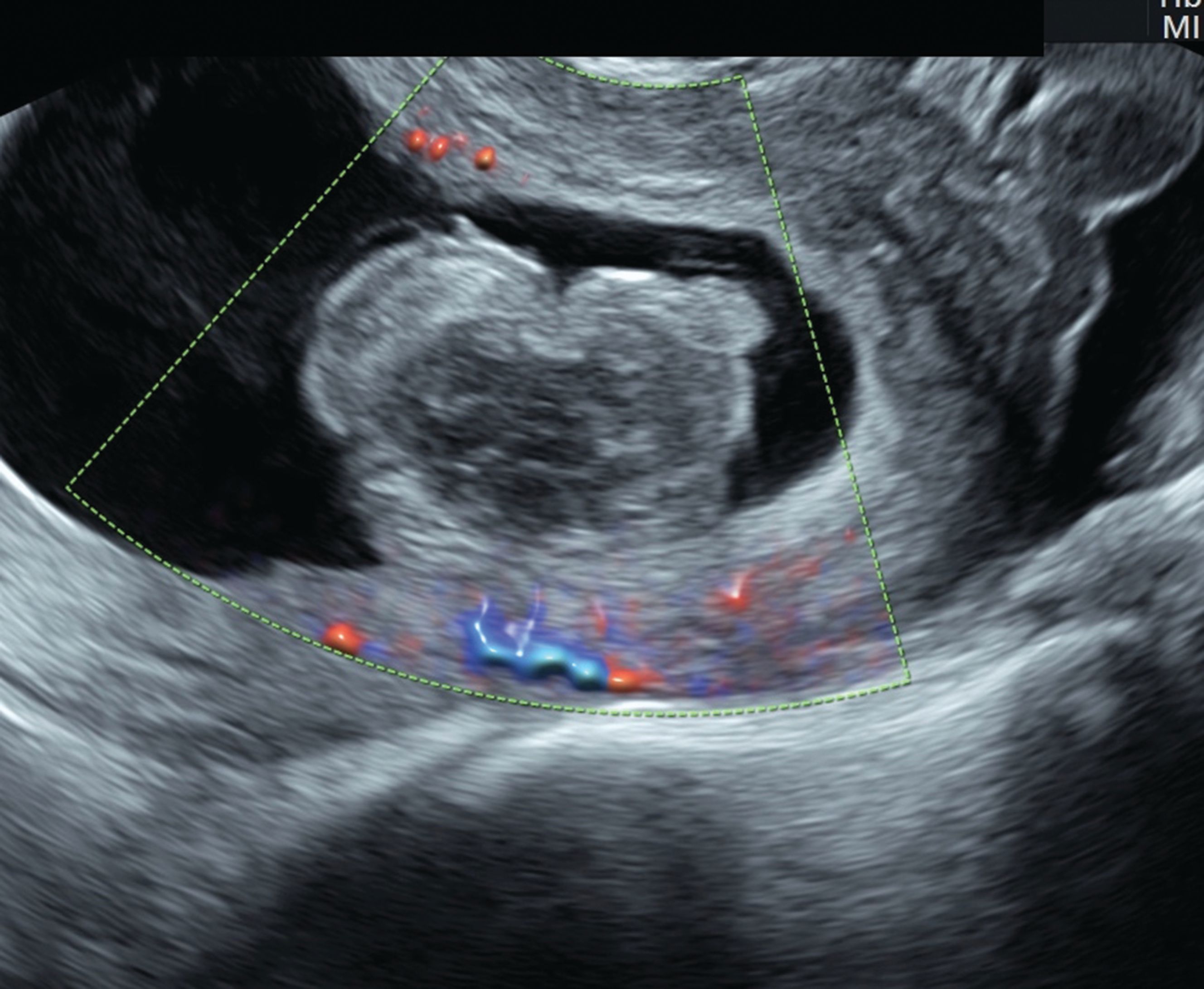
Normal And Abnormal Us Findings In Early First Trimester Pregnancy Review Of The Society Of Radiologists In Ultrasound 2012 Consensus Panel Recommendations Radiographics

Why To Avoid Keepsake 3 D And 4 D Ultrasounds Your Pregnancy Matters Ut Southwestern Medical Center
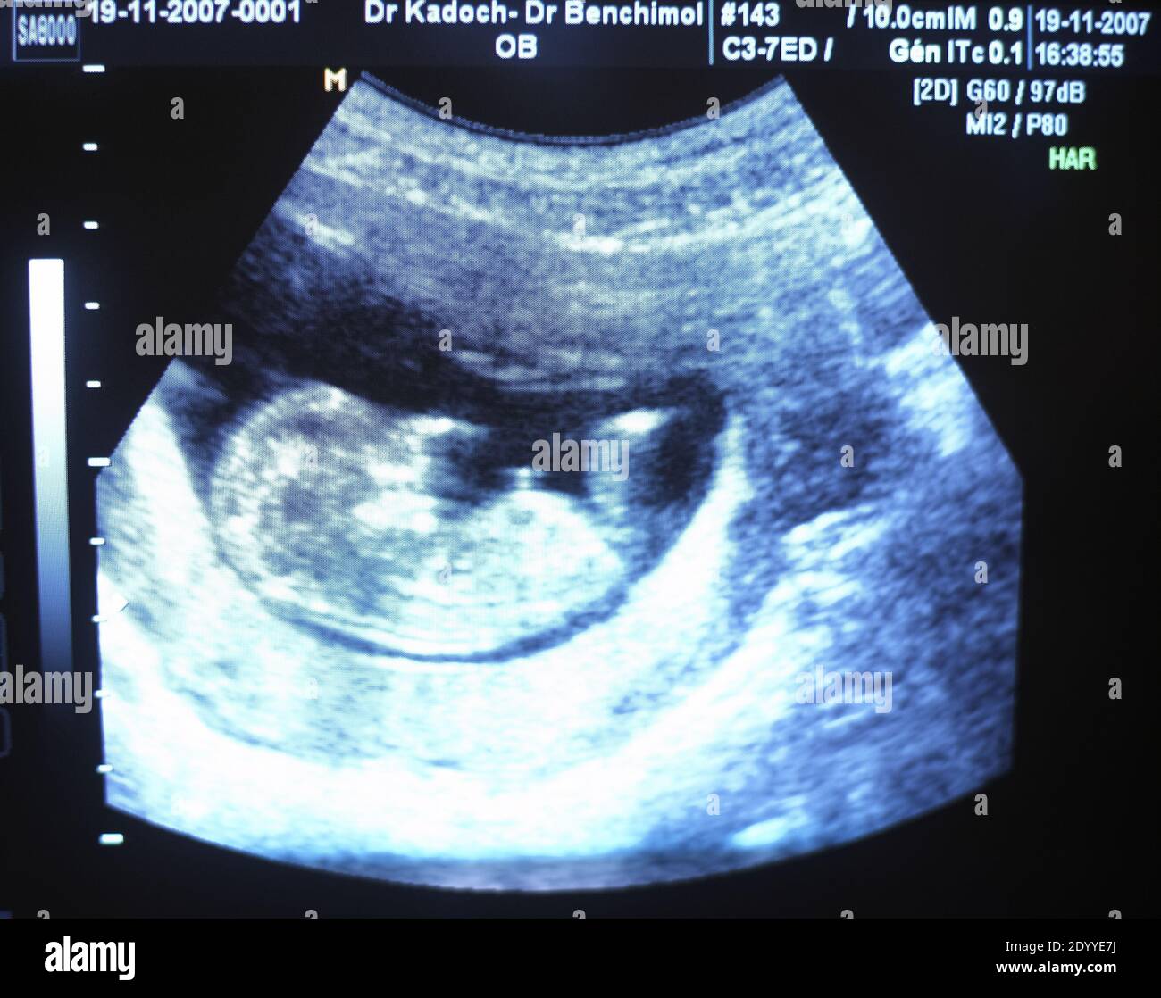
Week By Week Pregnancy Scan Photos Weeks 4 40 Netmums
12 Weeks Fetus Hi Res Stock Photography And Images Alamy

Sonographic Detection Of Fetal Abnormalities Before 11 Weeks Of Gestation Rolnik 2020 Ultrasound In Obstetrics Gynecology Wiley Online Library
An Imaging Approach To Early Pregnancy Failure
1st Trimester Ultrasound Scanning

Fetal Pictures Of Ultrasounds Gallery Imaging Technology News
What To Expect At An 8 Week Ultrasound Huggies Us

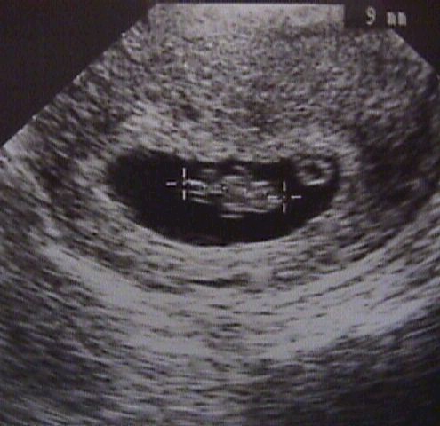
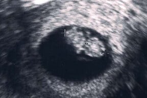
4 Week Ultrasound June 2015 Babies Forums What To Expect
5 Weeks Fake Ultrasound With Instant Download Baby Maybe
What To Expect At Your Six Week Ultrasound Appointment

456 Pregnancy Week 4 Images Stock Photos Vectors Shutterstock